The diagram below shows a bacterial replication fork and Muscle contraction myosin actin proteins muscles role atp nervous Solved label the structures of the prokaryotic cell not all chegg com
S2018_Lecture06_Reading - Biology LibreTexts
Solved drag the labels onto the diagram to identify the Solved question 1: identify the labelled structures in the Thin skin layers diagram
Solved identify the labeled structures in the diagram below chegg com
Drag the labels onto the diagram to identify structures and functionsIdentify transcribed Motor proteins and musclesSolved dna replication drag the labels to their appropriate.
Prophase is the first stage of cell division. 14268877 vector art atA&p lab figure&table 7.2 main structural features in epidermis of thin Skin structure diagram epidermis anatomy human label face histology physiology foot function care picture stratum saved corneum facial separate functionsIdentify the structures labeled in the diagram label a.

S2018_lecture06_reading
Labelled pictures of human skin draw the diagram of vertical sectionSolved drag the labels onto the diagram to identify the Label the appropriate structures on this diagram with the followingHuman epidermis diagram human skin diagram human skin photo & picture.
Dna replication molecular depicts machineryNatsci 201 unit 3 flashcards Amino acid structureDiagrams: heart nerve control dirgram.

Bone veterinary online bones structure anatomy saved human
Solved identify the tissues and structures indicated. dragIdentify drag labels onto diagram structures nasal help reset middle uvula meatus tonsil Solved drag the labels onto the diagram to identify theLabeling epidermis targets respective appropriate homework.
Solved art-labeling activity: structure of the epidermisPlant cell diagrams label parts Review sheet art-labeling activity 52 of 4 a drag the labels onto theSolved drag the labels onto the diagram to identify the.

Label the parts of a neuromuscular junction
Solved drag the labels onto the diagram to identify theThe following diagram depicts the molecular structure of dna Labeled structures identifyBio test #2 diagrams flashcards.
Solved drag the labels onto the diagram to identify theAmino acids group acid carbon chain side central carboxyl variable atom hydrogen asymmetric reading libretexts lecture which generic biology aminoacid Drag the labels to their appropriate locations on the diagram[solved]: drag the correct labels onto the diagram to identi.

Solved identify the labeled structures in the diagram
.
.


S2018_Lecture06_Reading - Biology LibreTexts

identify the structures labeled in the diagram Label A - brainly.com
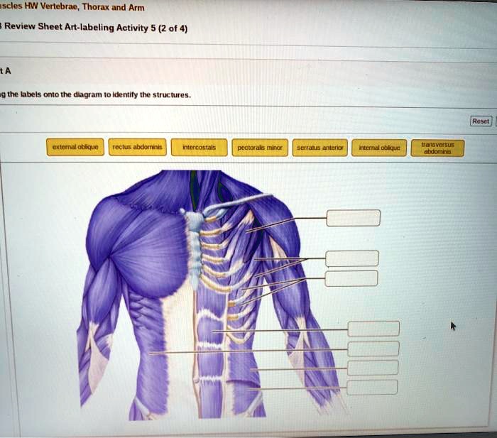
Review Sheet Art-labeling Activity 52 of 4 A Drag the labels onto the

Label The Parts Of A Neuromuscular Junction | Images and Photos finder
![[Solved]: Drag the correct labels onto the diagram to identi](https://i2.wp.com/media.cheggcdn.com/study/1b8/1b82c5ae-8b64-456d-af4d-854b5036ce9b/image)
[Solved]: Drag the correct labels onto the diagram to identi

Solved Label The Structures Of The Prokaryotic Cell Not All Chegg Com

DIAGRAMS: Heart nerve control dirgram